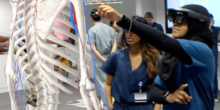
The HoloAnatomy® Software Suite* emerged out of a vision by CWRU and the Cleveland Clinic to establish a new Health Education Campus named the Sheila and Eric Samson Pavilion.
As building plans were made, interest in exploring the most advanced learning technology combined with an early introduction to Microsoft HoloLens led to a dramatic change in the CWRU School of Medicine (SOM) anatomy curriculum, from cadaver-based to a new digital, “Living Anatomy” curriculum. The result is a first-in-kind, engaging and academically tested and validated mixed reality medical anatomy education tool.
*patent pending.
Studies have shown that the HoloAnatomy® Software is not only effective and time-saving, but preferable over cadaveric dissection in some aspects.
In a controlled trial in the musculoskeletal system, students who learned using the HoloAnatomy® Software achieved the same scores as those that were in the cadaver lab, but the ones who learned in HoloLens did so in about half of the time. This has since been repeated and the results confirmed. Other studies have shown a dramatic improvement in students’ long-term retention of knowledge when using the HoloAnatomy® Software as compared to a cadaver lab.
The HoloAnatomy® Software has now been installed as the primary means of medical education at the CWRU SOM since the academic year 2019/2020, and was even modified to be taught virtually during the COVID-19 pandemic shutdown.
Learn more about AlensiaXR and the
HoloAnatomy ® Software Suite
PRESS
CWRU and IC HoloAnatomy App Partners Announced
CWRU announced that they will be partnering with five universities who have signed on to begin teaching with the HoloAnatomy®...

Study on HoloAnatomy published in Medical Science Educator
In this randomized controlled trial, we looked at the effectiveness of a mixed reality (MR) device - the Microsoft HoloLens...

Building the First Holographic Brain ‘Atlas’
Read more about the first ever 'atlas' of the pathways in the brain, visualized by the IC and developed in...
Study on HoloAnatomy Software published in Medical Education Journal
New study on the benefits of our HoloAnatomy® Software application as a supplement to traditional cadaveric dissection in medical education....
